Anatomy of the eye


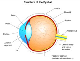
Structure of the eye ball: This is the cross-section(side view) of the eye. The eye works like the camera, transmitting light and images to the brain.
 Tear apparatus of the eye: Tears are formed in the Lacrimal Glands and the Accessory Lacrimal Glands. They are drained via the Lacrimal Ducts, at the nasal side of the eye, into the Nasolacrimal duct, which opens into the nose. That is why any eyedrops applied to the eye come into the nose and throat. Sometimes the Nasolacrimal duct may get blocked and it may then need to be openned surgically.
 Cross-section of the eyelid: The tarsal plate is like a backbone of the eyelid. It gives strength and flexibility to the eye lid. The muscles in the lid are attached to this structure. The Meibomian Glands are like sweat glands. It secretes a fatty substance which helps in maintaining the surface tension of the tears. A blockage of these glands leads to a condition called Chalazion.
 Cross-section of the Cornea: Cornea is the front transparent layer of the eye. It is a multi layered structure. The tears also form a thin layer on top of the cornea and protect and nourish it. A deficiency of tears can cause Dry eye. Shape of the Cornea is altered with LASER in Refractive Surgery(Surgery for correcting spectacle numbers). Any opacity or injury to this structure can cause permanent opacity and hence loss of vision.
 Division of the eye: The eye is divided in two main parts. The front or Anterior Segment. This has a water like fluid called Aqueous Humor.The back part or Posterior Segment has a thick jelly like substance called Vitreous Humor.
 Anterior Segment of the eye: It also has 2 sub-sections divided by the Iris. The Iris is like the shutter of camera. It controls the amount of light coming in the eye. The Aqueous Humor enters the chamber from the Ciliary Body and passes through the hole in the Iris called the Pupil, to exit in the angle between the Iris and Cornea. Any obstruction in the circulation of this fluid leads to rise in the eye pressure and can then cause a condition called Glaucoma.
 Lens of the eye: When this becomes opaque, it is called Cataract. Surgery is the only treatment for Cataract.
 Front view of the Retina: Retina is inner layer of the eye which when stimulated by light sends signal to brain and we perceive an image. The retina may get detached from the inner layer and is then put in its place by surgery. Diseases like Diabetes, High Blood pressure, old age, high myopia, etc can also damage the retina and can cause permanent loss of vision.
 Cross-section of the Retina: Retina is made up of 10 layers. Signal from here are carried by the Optic Nerve to the brain via the visual pathway. The Optic Nerve can be affected in many disease, like Glaucoma and Alcohol abuse.
 Eye Muscles: They control the eye movement. Any imbalance in the tone of these muscles can lead to Squint. Sometimes surgery is necessary to restore the proper balance.